Skip to Content

Three from Hopkins BME recognized at SPIE 2019

February 26, 2019
Left to right: Rohan Vijayan, Xiaoxuan Esme Zhang, Jeff Siewerdsen

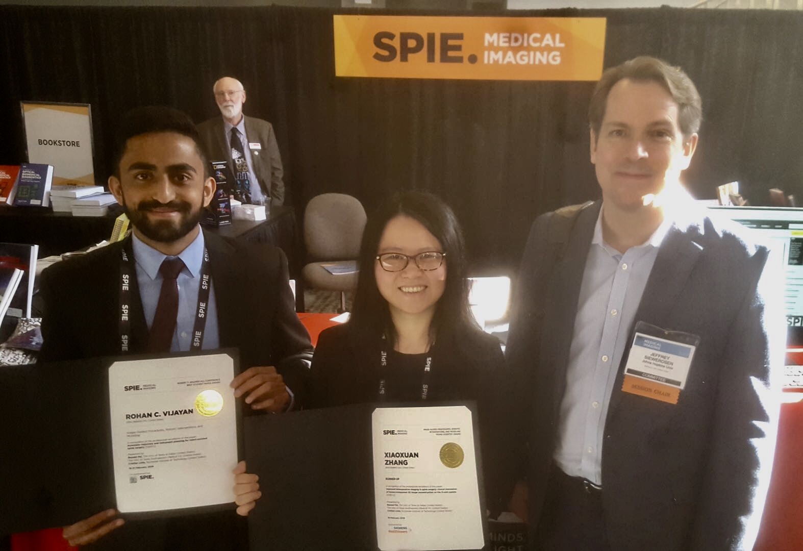
More than 1,200 people gathered in San Diego, CA last week for the annual SPIE Medical Imaging Conference, which featured more than 1,000 presentations, including three dozen involving Johns Hopkins researchers, on the latest advances in medical imaging. Three members of the Johns Hopkins Department of Biomedical Engineering were recognized at the conference for the impact of their research on the field. All are members of the I-STAR Lab, headed by faculty members Jeff Siewerdsen, Webster Stayman, and Wojciech Zbijewski.

Each year, the conference features cutting edge technology in areas such as digital pathology, image-guided procedures, computer-aided diagnosis, image processing, machine learning, and other biomedical applications.

Those recognized from Johns Hopkins BME include:

Wenying Wang, PhD student – Robert Wagner Best Student Paper, Winner

Wang is working to reduce the dose of potentially harmful ionizing radiation that patients receive while undergoing computed tomography (CT), a widely used imaging method for disease diagnosis and image-guided therapy. Wang used a new filtration scheme involving multiple aperture devices (MADs) to develop an exposure delivery design that reduces radiation dose while retaining image quality for single-organ imaging and other localized CT applications.

Rohan Vijayan, PhD student –Robert Wagner Best Student Paper, Finalist

Vijayan is developing an algorithm to automatically plan screw trajectories in CT scans of patients who are undergoing spinal surgery. These plans are designed to streamline surgical procedures and improve the accuracy of robot-assisted screw placement, providing near real-time quality assurance and enabling clinicians to better predict spinal surgery outcomes.

Xiaoxuan Esme Zhang, Research Scientist –Young Scientist Award, Runner-up

Zhang is working on a CT image reconstruction algorithm that improves visualization of patient anatomy and reduces radiation exposure during image-guided procedures. Working closely with physicians and industry collaborators, her team was the first to apply this algorithm to clinical studies, showing promising image quality results for patients undergoing surgical procedures to receive spinal implants.

Read the Johns Hopkins University privacy statement here.

Accept