
I-STAR Lab refines ultra-low dose CT scans
Computerized tomography (CT) has revolutionized the field of medical imagery, allowing doctors to peer inside patients as never before and leading to remarkable advances in diagnostics.
As good as those advances are, however, they are not perfect and come with certain risks to the patient. Researchers at the Imaging for Surgery, Therapy and Radiology (I-STAR) Lab in the Department of Biomedical Engineering are making great strides in improving CT image quality, while reducing the serious negative consequences of the technique.
The challenges are considerable. First, CT uses X-ray radiation, which is known in high doses and repeated exposures to cause cancer, among other negative health effects, particularly for patients who undergo multiple scans during treatment. Techniques that simply reduce dosage produce inadequate images. Accuracy can similarly be affected by metal devices, such as screws and plates implanted in a patient, compromising precision exactly where it’s needed most, at the site of investigation.
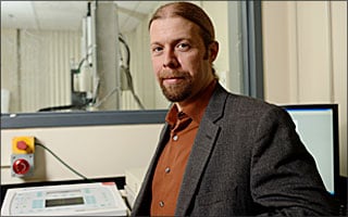
Researchers at I-STAR have applied both pragmatism and advanced computer modeling to make their improvements. “As good as CT is, it’s not a ‘smart’ technology,” says J. Webster “Web” Stayman, PhD, an assistant research professor in the Department of Biomedical Engineering’s I-STAR lab. “With most patients, though, there is a wealth of medical history and images that have been essentially left on the floor. We use these to make CT smarter.”
Such information, ranging from detailed CAD drawings of surgical implants and other devices to previously acquired patient images, helps inform Stayman’s techniques.
“If we know the exact dimensions of a screw, for instance, we can in effect use that data to fill in missing information, improving image quality and providing surgeons answers to important questions that would be hard to answer using traditional CT: Is that screw in the right place? Are there complications like fractures or bleeds?” Stayman says.
In addition, Stayman is perfecting new computer algorithms that reduce the number of images necessary for diagnosis, improving the speed and optimizing the methods used to collect data. Each of these techniques allows Stayman to lower radiation doses, minimizing risk to the patient — maybe the best improvement of all.
— Andrew Myers