
Huge Hopkins presence at SPIE Medical Imaging symposium
The annual SPIE Medical Imaging symposium held in Houston, TX featured 14 talks from faculty and students from the Johns Hopkins Departments of Biomedical Engineering, Radiology, Neurosurgery, and Orthopaedic Surgery.
Martin Pomper, professor of radiology, gave a keynote presentation on biomedical applications in molecular, structural, and functional imaging. Others selected for distinguished presentations were Alisa Brown, an undergraduate student majoring in biomedical engineering, and Thomas Yi, a research assistant working in the Johns Hopkins Carnegie Center for Surgical Innovation.
Yi presented his work on x-ray-guided robot-assisted spine surgery, describing how he used 3D2D registration to automatically position a robotic drill as a guide for high-precision placement of spinal pedicle screws. His work goes beyond conventional approaches to robotic assistance in a way that is more accurate in the presence of deforming anatomy, and operates without additional optical markers and surgical trackers.
Brown presented on the design and validation of a large, open-source library of rigid-body markers for surgical navigation. Her work yielded a library of dynamic reference frames that could be produced using a 3-D printer, and facilitates research and development in surgical navigation using multiple tracked tools.
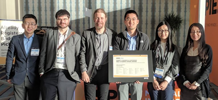
Andrew Mao, a master’s student working with assistant professor Webster Stayman, was awarded Best Student Paper within the Physics in Medicine category, as well as the All-Conference Robert F. Wagner Award.
Other presentations from Johns Hopkins affiliates covered topics such as an algorithm for cone-beam CT neuro-angiography, a model for spatial resolution and noise in flat-panel detector cone-beam CT images, a method for selecting kay parameters in prior-image-based 3-D image reconstruction, and curved detector designs in compact geometry CBCT systems.
- PhD student Runze Han, who described a method for automatically planning surgical approach in pelvic trauma surgery
- Research scientist Niral Sheth, who reported on the imaging performance of an intraoperaticve C-arm incorporating a high-performance CMOS x-ray detector
- Postdoctoral fellow Ali Uneri, who reported findings from his work using model-based image reconstruction on the Medtronic O-arm
- PhD student Pengwei Wu, who presented on the “Reconstruction-of-Difference” algorithm for cone-beam CT neuro-angiography
- PhD student Wenying Wang, who described a model for spatial resolution and noise in flat-panel detector cone-beam CT images reconstructed using penalized likelihood estimation
- Postdoctoral fellow Hao Zhang, who presented a method for prospectively selecting key parameters in prior-image-based 3-D image reconstruction in a manner that reliably controls the influence of new and prior image information in MBIR
- PhD student Qian Cao, who reported on the high-resolution characteristics of a CMOS detector in cone-beam CT for imaging of bone micro-architecture
- Research associate Alejandro Sisniega, who described the influence of curved detector designs in compact geometry CBCT systems, including the importance of antiscatter grid selection and bowtie filter design.
- Postdoctoral fellow Michael Brehler, who reported on methods for quantitative analysis of trabecular bone structure in high-resolution cone-beam CT
- PhD student Michael Ketcha, who described a mathematical model for image registration performance that includes not only the influence of image quality (noise and resolution), but also the confounding influence of soft-tissue deformation.
- Undergraduate student Benjamin Ramsey, who presented a variation on atlas-based registration in which sub-atlases are used in a series of stages according to similarity in principal component analysis to guides more accurate deformable registration
- Postdoctoral fellow Tharindu De Silva, who reported on a fast slice-to-volume image registration method for ultrasound imaging in spinal interventions
The annual SPIE Medical Imaging conference focuses on the latest innovations related to underlying fundamental scientific principles, technology developments, scientific evaluation, and clinical applications. The symposium covered a wide range of medical imaging modalities including medical image acquisition, display, processing, analysis, perception, and decision support.