
CardioViz: Improving the treatment of tricuspid valve disease with 4D fusion cardiac imaging
- Vivek Gopalakrishnan
- Christine Ji
- Angela Lu
- Bolaji John
- Chester Huynh
- Ethan Levy
- Lauren Pan
- Zan Chaudhry
- Rani Hasan, MD, MHS
- Elizabeth Logsdon, PhD
- Jeffrey Siewerdsen, PhD
- Mathias Unberath, PhD
- Jon Resar, MD
- Stefan Zimmerman, MD
- Taylor Holt
Abstract:
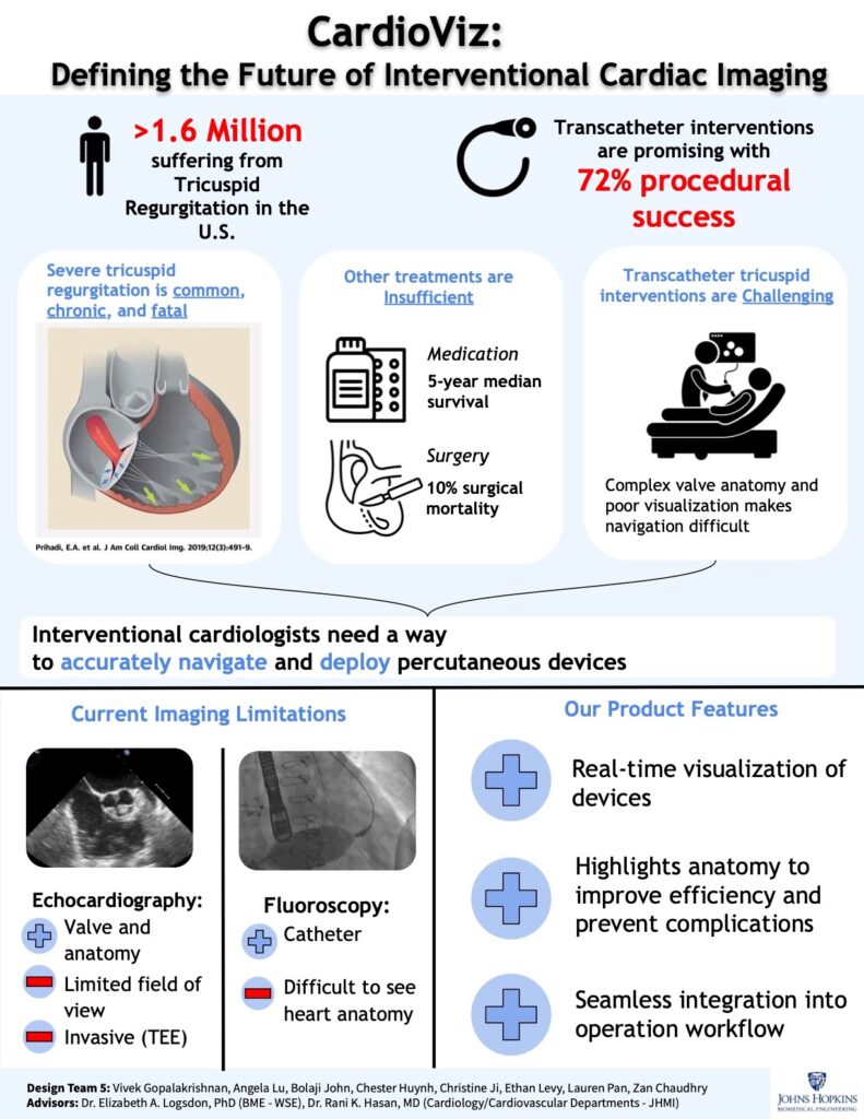
Approximately 8 million patients across the United States and Europe suffer from moderate-to-severe tricuspid regurgitation (TR), a chronic and progressive disease that increases risk of arrhythmia, heart failure, and mortality. Patient options for managing the disease are insufficient as medication merely treats symptoms and has low long-term effectiveness, and tricuspid valve surgery entails the highest surgical mortality rate of any open heart procedure. While minimally invasive transcatheter aortic and mitral valve interventions have revolutionized the treatment of structural heart disease, these advances have yet to translate to the treatment of tricuspid valve disease. A combination of complex tricuspid valve anatomy and poor intraoperative visualization limits the ability of interventional cardiologists to accurately deploy percutaneous devices as anchor points in the tricuspid valve apparatus. Thus, there is a critical need for effective transcatheter interventions as they are uniquely poised to be a safer, minimally invasive alternative to open heart surgery. To improve visualization for tricuspid procedures, our team is developing a 4D image fusion system that combines and annotates multiple cardiac imaging modalities in real time, providing clinicians an augmented view of a patient’s heart during procedures.