Sharper images with MRI

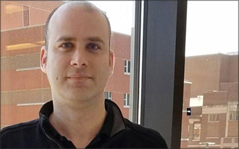
At the Johns Hopkins Dynamic Imaging Laboratory, Daniel Herzka and colleagues are pushing the boundaries of magnetic resonance imaging.
“MRI is a wonderful camera,” explains Herzka, a BME assistant professor. “It allows us to see into the human body. Unfortunately, it’s also very sensitive, particularly to motion.” As physicians seek images with higher and higher resolution to examine smaller and smaller structures, scans take progressively longer, he says, and patients in the machine tend to move more. “It’s natural for patients to shift their feet, move their heads, swallow, or cough, all of which can interfere with imaging, especially when looking at really small things.”
Herzka’s group modifies MRI machines, altering both how the information is acquired and the images are reconstructed. In the lab, located on the School of Medicine campus, they work to use MRI to see organs better. As part of that, they develop techniques to compensate for many types of motion such as breathing when imaging the heart or slight shifts in position when imaging knee cartilage. They also work on imaging faster so that motion doesn’t matter.
The efforts are critical in areas like heart disease, where Herzka is working to enable better visualization of scar tissue that forms after an injury like a heart attack. With very accurate knowledge of the location of scar tissue, physicians can design better treatments for cardiac arrhythmias. Herzka’s group is also tackling imaging of the inner ear, nerve roots that cause pain, and thin layers of cartilage in joints. In each of these projects, the goal is to see something that can’t be seen well with current clinical systems.
Additionally, Herzka’s group uses a quantitative approach, developing new methods to measure physical parameters of tissues with high accuracy. “Though MRI produces images, it can also be used to measure basic properties of tissue, giving us one more way to understand a disease process,” he says. “Seeing with more clarity into the human body is invaluable throughout medicine.”
Besides working with cardiac electrophysiologists, Herzka collaborates with musculoskeletal radiologists and neuroradiologists, always pushing to see smaller and smaller structures. “By showing physicians what they couldn’t see before, our images and measurements can change the trajectories of patients with complex disease.”
“I’m interested in getting as much as we can out of these systems and getting the best possible images with the most diagnostically relevant information,” Herzka says. “I’m particularly attracted to open problems, where imaging has been deemed unfeasible and the common thought is, ‘You can’t see that with MRI.’ Sometimes you can’t; it takes too long or it’s just too difficult for our current technology. So we develop new imaging methods so that sometimes you can, which is great. We may just have to work a little harder to see deeper and better.”
— Karen Blum
