Skip to Content

Scot Kuo to share $17 million Chan Zuckerberg Award

March 19, 2019

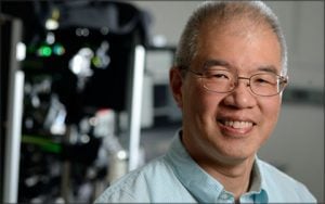
Scot Kuo, Ph.D. makes the super small visible. He tracks how cells—often smaller than the width of a human hair—move and interact with other cells. He uses high-resolution microscopes to zoom in on individual parts and proteins within cells, down to the molecular level. Kuo’s pioneering work to improve the field of microscopy has helped hundreds of scientists at Johns Hopkins look far more closely at cells and the structures within them.

Now, Kuo is one of 17 scientists in the U.S.—engineers, physicists, mathematicians, computer scientists and biologists—who will share in $17 million from the Chan Zuckerberg Initiative (CZI), the foundation launched by Facebook founder and CEO Mark Zuckerberg and his wife, pediatrician Priscilla Chan.

Kuo, associate professor of biomedical engineering and cell biology at the Johns Hopkins University School of Medicine, will receive $1.2 million over the next five years. He is also director of the School of Medicine’s Microscope Facility and director of imaging at the Institute for Basic Biomedical Sciences.The Microscope Facility is outfitted with 21 high-powered microscopes that vary in their ability to see fine detail and the speed of images captured. Most of the microscopes in the facility work like the Google Earth of biology, where scientists can zoom in and out of cells and other biological samples.

Kuo has spent more than 20 years taking existing technologies and tools and improving them—with better software, hardware and sometimes a roll of duct tape—to help his scientific peers answer the most pressing questions in biology. The CZI funds will help Kuo make new inventions that will push the field of microscopy further and spur scientists to make progress toward improving human health.

“Although optical and electronic hardware play critical roles for enabling new imaging technologies, it’s the software that often determines the usability of the equipment for biological and biomedical research,” says Kuo.

Physicists have a strategy to solve a similar problem in astronomy where wind and other atmospheric elements distort the image of stars so that they appear to blur and twinkle. Kuo says that telescope technology, called adaptive optics, is being applied to biology, too. He and other scientists are developing algorithms to map and account for how light bends in tissues so that they can sharpen biological images.

A kidney slice is shown from a microscope.
A kidney slice from a mouse with polycystic kidney disease, taken from a microscope in Scot Kuo’s facility. The image has subcellular resolution that is powerful enough to monitor chemical changes inside the cell caused by drugs used to treat the disease. Credit: Scot Kuo

Read the full list of newly designated CZI imaging scientists.

Category: Faculty
Associated Faculty: Scot C. Kuo

Read the Johns Hopkins University privacy statement here.

Accept