Skip to Content

Scot Kuo: BME’s gadget man

April 15, 2016
Scot Kuo

A biochemist and cell biologist by training, Biomedical Engineering Associate Professor Scot Kuo made a name for himself developing technologies to better understand the mechanical functions of cells. These include optical tweezers, a tool to measure forces between and within molecules, and laser-based nano-tracking, an instrument that helped demonstrate that the bacterial pathogen Listeria moves in a jerky, steplike fashion when it infects cells.

But when he got the opportunity to direct the Johns Hopkins School of Medicine’s Microscope Facility in 2006, he couldn’t resist. The position calls on a number of his interests — like advising faculty on what tools can best help them achieve desired research results or suggesting good materials and design strategies to biomedical engineers looking to create their own equipment.

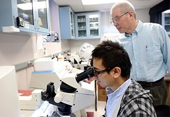
A student looks into a microscope as a faculty member stands behind him.

“I really enjoy talking to people and troubleshooting,” he says, as well as connecting researchers with others using technology that could be shared for mutual benefit. “I feel like a social butterfly pollinating different ideas. It’s a lot of fun to be at that nexus.”

With his prowess writing grants for federal funds to purchase new equipment (his success rate is 80 percent), Johns Hopkins in January became the third institution in the world to acquire a lattice light sheet microscope. The device, invented by Nobel Prize winner Eric Betzig in 2014, allows rapid collection of high-resolution fluorescence images under a very low light level, which minimizes the cell damage that often limits the duration of experiments. It records three-dimensional movies of molecules, cells, and embryos in fine detail — like an immune system T cell attacking another cell — over longer periods than was previously possible with fluorescence, at a speed up to 1,000 frames per second.

Interest in the microscope has been extremely high, says Kuo. When he gave an introductory talk about the device at the School of Medicine last October, the subject “had people spilling out onto the floor.”

Kuo and his staff of five also support two other new purchases — a high-pressure freezer with optogenetics features and a high-content imager — in addition to about 18 other microscopes.

The high-pressure freezer allows quick freezing of tissue samples to minus 196 degrees Celsius and won’t form ice crystals, allowing investigators to use chemicals to manipulate the cells while preserving their structural properties. This device can flash light on nerve or other tissue to trigger action potentiallly milliseconds before it’s frozen, enabling the study of dynamic changes within excitable cells.

The high-content imager, which will incorporate a robot, can quickly scan specimens in large microtiter plates, recording up to 6,000 pictures of nearly 400 samples in a half-hour. It’s a huge time and work savings over the traditional methods of preparing each specimen separately and taking one picture at a time, Kuo says.

“Keeping an eye on all the upcoming technologies, there’s plenty of microscopes that we don’t have yet here,” he says. He remains on the lookout for the ones that will best benefit the Johns Hopkins scientific community.

Category: Research
Associated Faculty: Scot C. Kuo

Read the Johns Hopkins University privacy statement here.

Accept