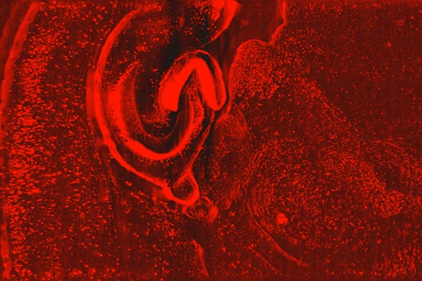
A team of researchers from Johns Hopkins, including Assistant Professor Ji Yi and Professor Patrick Kanold, have shown that a new microscopy technique can capture dynamic 3D images of an entire zebrafish larvae while maintaining cellular resolution in all three dimensions. By giving scientists an unprecedented view of how cells interact in their most natural state, the new technique could provide information that is used to develop new treatments for diseases, for example.
“Unraveling underlying cellular structures and their interaction is fundamentally important to understanding life,” said research team leader Yi. “However, the limitations of light diffraction make it difficult to image with 3D cellular resolution over large areas of several millimeters. We circumvent the trade-offs between field of view, depth resolution and imaging speed to achieve 4D cellular resolution over a much larger field of view than previously possible.”
Learn more on Optica.

