
Promising new imaging solution for traumatic brain injury and intracranial hemorrhage
The ability to detect intracranial hemorrhage (ICH) with an imaging technology suitable to the point of care could offer significant advances in applications ranging from image-guided surgery (IGS) to diagnosis of stroke or traumatic brain injury (TBI).
For example, TBI is estimated to result in 1.7 million visits to emergency rooms each year and is a contributing factor in 30% of injury-related death in the US. Accurate diagnosis of acute TBI is extremely important in determining the appropriate course of treatment and would greatly benefit from availability of imaging at the point-of-care (e.g. ambulance, ICU, or urgent care center) or even directly in the environment where TBI occurs (e.g. athletic or war theater).
Currently, multi-detector CT is the front-line method to determine fresh blood hemorrhage in the brain, while MRI is used extensively for evaluation of subtle diffuse lesions in more chronic stages of pathology. However, neither multi-detector CT nor MRI are particularly well suited for point-of care imaging due to relatively high cost, large footprint, and limited portability.
In recent years, flat-panel detector-based cone-beam CT (FPD CBCT) has emerged as a promising technology. More portable due to weight and size, this imaging method could provide a valuable means for point-of-care imaging. However, soft-tissue imaging of the brain with the current generation of FPD-CBCT systems is challenged by poor contrast resolution of soft tissues, brain, and blood, along with lower image uniformity, higher noise, and a higher level of artifacts.
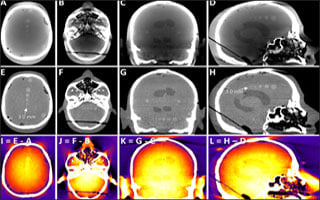
To advance the image quality of cone-beam CT to a level suitable for soft-tissue brain imaging, a Johns Hopkins research team that includes the Department of Biomedical Engineering Professor Jeffrey Siewerdsen and researchers in the I-STAR Lab in collaboration with clinical experts in Neurology Dr. Vassilis Koliatsos and Radiology Dr. Nafi Aygun have developed a comprehensive framework for artifact, detector lag, veiling glare, and beam hardening correction.
Recently, Dr. Alejandro Sisniega, a postdoctoral research fellow in the I-STAR Lab, completed work in a landmark paper reporting high-quality imaging of head injury with CBCT. The Johns Hopkins research team revealed that the correction method yielded improved image quality sufficient for reliable diagnosis of subtle intracranial hemorrhage. The results serve to further motivate development and translation of a dedicated system for this application, now underway in academic-industry partnership.