
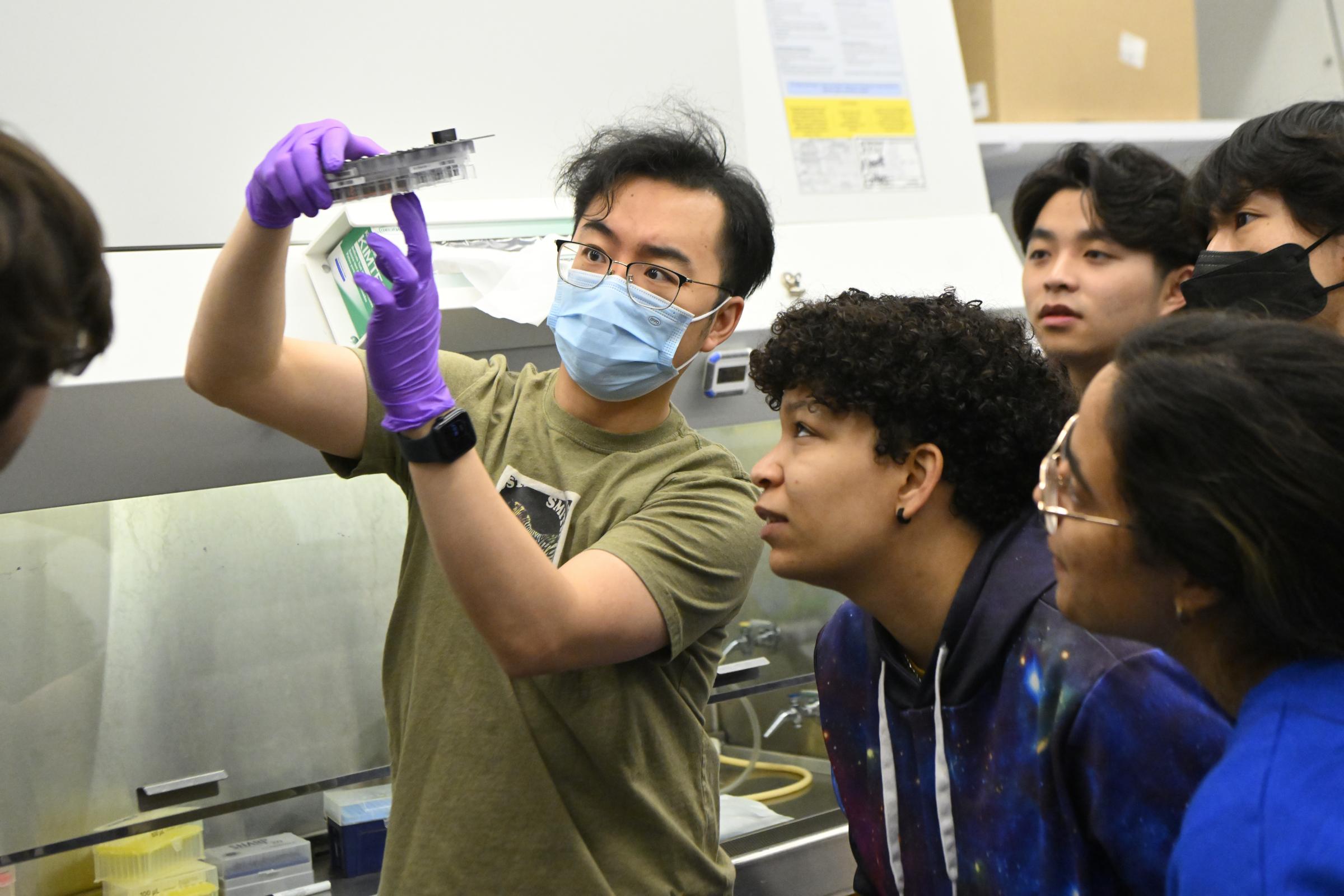
On a recent Tuesday, biomedical engineering students gathered in Clark Hall to see the fruits of their labor. After spending the better part of two weeks coaxing human induced pluripotent stem cells, or iPSCs, to develop into cardiac muscle tissue by adding biological factors to the cells in a culture plate, students met to measure the force of contractions in the tissue when an electrical stimulus is applied.
“Oh my gosh, it’s there!” one student exclaimed when seeing the twitching tissue, a miniaturized model of what happens when someone flexes their arm.
This work was part of a new course called Microphysiological Systems, created by Deok-Ho Kim, a professor of biomedical engineering. Kim says the curriculum for the class, the first of its kind at Johns Hopkins, focuses on applying biological and engineering fundamentals to design microphysiological systems, or MPS, such as organ and tissue chips, 3D-printed tissues, and organoids—artificially-grown masses of cells that resemble an organ.
MPS are used to study human disease, drug development, and precision medicine. Kim created the course because he believes that learning to work with MPS is essential for the next generation of biomedical engineers and scientists.
“MPS are now and will continue to be an integral part of advancing medical science and drug development, both for fundamental discoveries and testing clinical therapies,” Kim says. “My course introduces MPS and provides hands-on experience in the design, construction, and implementation of MPS platforms.”
The class features lessons on human stem cell technologies, organs-on-a-chip engineering, and education on MPS models for the heart, brain, lung, gut, kidney, tumors, and vascularization. The course also covers tissue chips for space biology, regulatory tools, and industrial applications.
The laboratory portion offers students hands-on experience in MPS design and application. Students learn biofabrication techniques such as microfluidics (manipulation of microscale fluid flow), microfabrication (making of miniaturized structures), and 3D bioprinting to create in vitro miniaturized, 3D complex human tissue models. In addition to the skeletal muscle work, students also learn about iPSC maintenance, turning iPSCs into heart tissue, and using MPS for cancer drug screening.
Kim has developed several MPS models and is a principal investigator on a National Institutes of Health-sponsored grant to study a muscular dystrophy tissue-on-a-chip model for future clinical trials. Kim also has twice sent human heart tissue-on-a-chip specimens to the International Space Station to study the effects of microgravity on heart cells’ mitochondria (power supply) and their ability to contract. In 2015, he launched a company, Curi Bio, which develops human MPS platforms for drug discovery.
