Skip to Content

Learning the science behind imaging devices

May 18, 2017
Student gather around a table to watch a demonstration.

Johns Hopkins students in the upper-level Imaging Instrumentation course showed off their year-end projects in late May in the BME Design Studio. Taught by Web Stayman, an assistant professor in the Department of Biomedical Engineering, the undergraduate and graduate students demonstrated projects that ranged from a system to replicate an endoscopic procedure to a method of measuring blood flow in the body.

“I was excited to see the students leveraging all of the hands-on experience they have gained throughout the semester, and applying those skills in final projects of their own design,” says Stayman. “This year, the class had four excellent imaging system designs developed, constructed, programmed, and optimized using knowledge from the first-half of the class.”

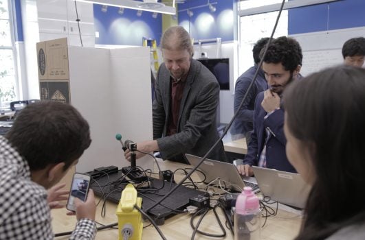
Throughout the semester, the students gained hands-on experience with the characterization, integration, data processing, and evaluation of imaging systems. After undertaking a series of labs for the first half of the course, they applied what they’ve learned to complete their own team projects. In the BME Design Studio in Clark Hall, teams presented designs to an audience of students and faculty.

Team one has a device aimed at a small ball on the table.

Team 1: Object Detection & Tracking

The team of four students created an object detection algorithm to program a small camera to automatically follow a designated object as it moved back and forth along a track. Using background subtraction, the camera was able to detect the object’s movement, eliminate the white backdrop, and produce a live video feed.

This device looks like a hallow black box with wire inside.

Team 2: Photometric Stereo Imaging for Endoscopic Topography

Using a commercial endoscope and a series of LED lights, the team constructed a photometric stereo setup to create a 3-D image. Variable angular illumination was achieved by controlling individual LEDs. The sequence of illumination data was processed to form 3-D height maps of various objects including hemisphere targets, a molded playdough object, and a colon phantom to replicate an endoscopic procedure.

This device has a bird squish toy and a phone inside a black box.

Team 3: 3-D Model Reconstruction and Texture Mapping Based on Structure from Motion

Students worked to create a system that can produce a digital 3-D model using a setup that included three LED light panels, a rotating stage, and a camera. A series of 72 images taken from different angles was captured and used to create a 3-D model. Details of the image were enhanced using texture mapping.

A student puts his hand inside a black box with a red light.

Team 4: Laser Speckle Flow Imaging

Speckle imaging refers to a random, granular pattern that is captured when light is reflected and scattered off an object. The team of students designed an apparatus to measure the blood flow of a member’s finger. The control image showed a typical speckle signal, while the image taken when a blood pressure arm cuff was activated on the subject’s arm displayed a much lower speckle signal, representing a decrease in blood flow.

Learning the intricacies of imaging devices has given these students an understanding of the physics, engineering, and algorithms that are needed to create these systems, Stayman says, and will prepare them for future research, careers or other pursuits outside the classroom. This course is offered to undergraduates (580.493) and graduates (580.693) every spring.

Read the Johns Hopkins University privacy statement here.

Accept