
Engineers at Johns Hopkins have solved the problem of distorted imaging scans that plague surgeons who need to use them to assess the placement of metal implants. They have developed a simple and practical solution to “steer” a scanner and direct its imaging beam so that most of the distortion problem is remedied.
Metal implants are commonly used to stabilize bones and structures in the body. During an operation, surgeons use a C-arm X-ray or CT (computed tomography) scanner to see if an implant is correctly positioned, has caused hemorrhaging or is impinging on adjacent nerves.
Problems with the implant can necessitate second surgeries that are expensive and often have poor outcomes, say the Johns Hopkins engineers.
“Metal artifacts are a giant headache for surgeons,” says Jeffrey Siewerdsen, professor of biomedical engineering at Johns Hopkins and leader of the team which published results of its proof-of-principle concept in the May 19, 2020, issue of the journal Physics in Medicine & Biology. “The beauty of the system we developed is that it has minimal change in the operating room workflow, time and cost.”
Metal transmits X-rays differently than tissue or bone, often yielding distortions, or artifacts, which appear on imaging scans. Artifacts may make the implant appear larger than real and form streaks throughout the image, blocking the surgeon’s ability to see potential problems.
Currently, scanners have built-in systems to analyze the X-ray signals and reduce the impact of metal artifacts. However, Siewerdsen says, these systems are not very effective, and the artifacts are worse with larger and denser implants.
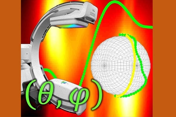
“We steer the C-arm scan to give data that contain the fewest errors that could cause image artifacts,” says Siewerdsen. “In some cases, the solution is as simple as tilting the C-arm.”
Using cadavers, torso models and surgery simulations, Siewerdsen’s team developed a computer algorithm that assesses the location of a metal implant and determines the best position of the CT scanner to avoid artifacts. They dubbed the method “metal artifact avoidance,” since it avoids errors in the original data, unlike other techniques that try to correct them later.
The researchers found that their tests of the algorithm and tilting of the C-arm reduced the magnitude of errors in the X-ray data and resulting artifacts in the image. In one example, a 5-millimeter-diameter metal screw was implanted into the spine of a cadaver. Without the metal artifact avoidance algorithm, the image of the screw was distorted so that it appeared to be 8–12 millimeters in diameter. With the algorithm, the screw appeared as its real size.
“The key is to compute a scan orbit in a simple way that can be easily run by the C-arm,” says Siewerdsen. “We were surprised at how well it works.”
To enable the computer algorithm to work on most C-arms, manufacturers would need to calibrate their scanners to be tilted in the operating room, notes Siewerdsen. He adds that the metal artifact avoidance method can be paired with different algorithms that correct other kinds of artifacts.