Diagnosing cancer is no easy game. When a doctor detects a possible tumor, a biopsy is ordered. Biopsies, however, are invasive and not very precise, and the evaluation requires the sample be sent out, sliced, stained, and studied under a microscope.
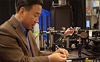
“We’re trying to put the microscope inside the body,” says Department of Biomedical Engineering Professor and Electrical and Computer Engineering researcher Xingde Li. Li is on the trail of revolutionary technologies at the cross-section of medicine and imaging, a field known as biophotonics.
“We created a very thin scanning fiber optic multiphoton microscope that goes inside the body, right to the place of interest, to help diagnose disease immediately, less invasively, and without any staining or processing,” says Li.
Li’s prototype is helping doctors to do remarkable new things. In one of many dramatic examples, Li’s endomicroscope, as he calls it, is being used to assist in brain surgeries.
“When you’re removing a brain tumor, you want to take as little of the healthy tissue as possible and as much of tumor tissue as possible. Our technology can help doctors see, in real time, where to cut and, more importantly, where not to cut,” Li explains.
Another exemplary application is to directly visualize the collagen fiber network in the cervix, from which the mechanical strength of the cervix, and thus the risk of preterm delivery, can be assessed.
The endomicroscope’s optical fiber is the same sort used to transmit high-speed data. The fibers are anywhere between a half to two millimeters in diameter. The fiber is flexible as well, so it can be guided to the exact spot needed.
Light travels down the fiber, which acts as a sort of flashlight, illuminating the tissues inside the body. The light reflects off the tissues (or is generated from the tissue such as fluorescence light) and is re-collected by the very same fiber, where it zips back through a computer to a waiting video monitor.
“We do it all noninvasively with a single ultrathin and flexible fiber endomicroscope in real time. That’s the real beauty of it,” Li says.
— Andrew Myers

