Skip to Content

3D nerve maps pinpoint the path to precision bone repair

November 3, 2025

The tiny nerves inside your bones—which are vital for maintaining bone strength and healing fractures—are finally being brought to light. These delicate structures have historically been incredibly hard to see and study, but Johns Hopkins University researchers have cracked this imaging challenge.

The team created an automated 3D method using artificial intelligence to precisely map the peripheral nerve network in dense bone tissue. Described in the journal Bone Reports, their machine learning-powered discovery is set to accelerate the search for new treatments for fractures and degenerative diseases like osteoporosis.

“The new method represents a significant leap forward because much research in the field still relies on manual tracing—which is both extremely tedious and subjective. By automating this process, we can better compare the findings between different labs and collectively unlock new research directions that weren’t available before,” said Warren Grayson, a professor of biomedical engineering and the study’s senior author.

Previously, researchers had to sit down and painstakingly trace individual nerve lines in massive datasets using a computer mouse. In contrast, the new, semi-automated method allows scientists to measure the entire nerve network quickly and accurately. This efficiency is critical for studying exactly how these nerves influence bone aging and healing, says Grayson.

To achieve this, the team developed a three-step system. First, a high-powered camera captures detailed 3D pictures of the bone. The main innovation was integrating a machine learning (ML) program called Ilastik®. The second step utilizes machine learning to sift through dense bone tissue to map and isolate the delicate nerves. This process effectively strips away all the distracting background noise, which is exacerbated by inflammatory conditions. Finally, a separate tool named Imaris analyzes this clean data to precisely calculate structural measurements such as nerve density.

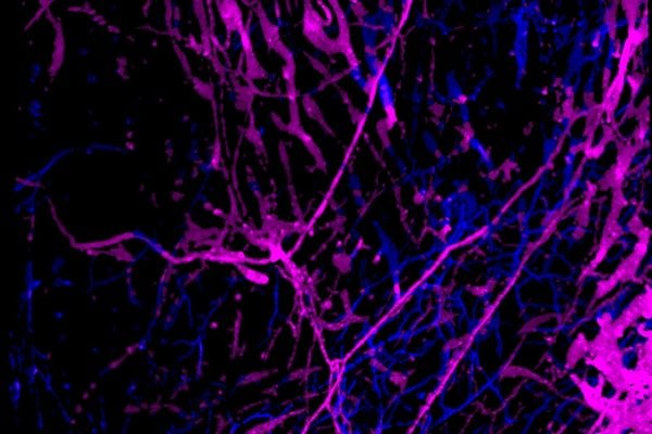
The method was successfully tested across multiple bone types, including the skull, the long bones in the foot (metatarsal bone), and the knee.

The need for better 3D nerve imaging became clear in the team’s previous study, which showed a significant change linked to aging: bones of the skull suffered a massive nerve loss, with approximately 50% of nerves disappearing in older mice.

“As you age, it’s pretty well known there’s an increase in bone fragility and fractures, and we don’t totally understand what mechanism drives that,” said lead author Allison Horenberg, a biomedical engineering PhD student. “Is the loss of nerves in the bone associated with those changes in fragility? That’s what this method is going to let us investigate in future studies.”

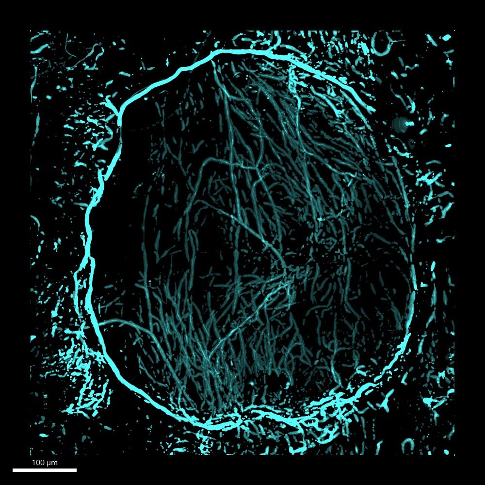
In ongoing studies, the method was applied to samples of injured skull bone to map nerves, blood vessels, and bone-forming cells after injury. The team hopes the detailed data from these nerve maps will finally help solve the mystery of why some bone injuries heal and others don’t.

Nerves in a non-healing skull defect four weeks after injury
Nerves in a non-healing skull defect four weeks after injury (Image Credit: The Grayson Laboratory)

The ultimate goal of the research is clinical: to apply these findings to tissue-engineered constructs—biomaterial scaffolds designed to regenerate bone in non-healing injuries, such as those resulting from blast trauma or cancer resections. Precise nerve data can help identify specific materials that should be added to these scaffolds to promote new bone growth. These next-gen bone grafts aim to outperform traditional grafts (which require harvesting bone from another part of the patient’s body) by eliminating the need for multiple surgeries and ensuring the new grafts are perfectly shaped.

“We’ve created this powerful new tool for science and lab research, but the potential is huge,” said Horenberg. “It means we can start asking questions we couldn’t before—like how nerves influence bone behavior during aging, injury, and disease and potentially help to identify novel therapies. That capability is going to speed up how quickly we can get effective new treatments into the clinic.”

Category: Research
Associated Faculty: Warren L. Grayson

Read the Johns Hopkins University privacy statement here.

Accept